Click image to see more details
Product Info Summary
| SKU: | PB9036 |
|---|---|
| Size: | 100 μg/vial |
| Reactive Species: | Human |
| Host: | Rabbit |
| Application: | IHC, WB |
Customers Who Bought This Also Bought


Product info
Product Name
Anti-LIF Antibody Picoband™
SKU/Catalog Number
PB9036
Size
100 μg/vial
Form
Lyophilized
Description
Boster Bio Anti-LIF Antibody Picoband™ catalog # PB9036. Tested in IHC, WB applications. This antibody reacts with Human.
Storage & Handling
Store at -20˚C for one year from date of receipt. After reconstitution, at 4˚C for one month. It can also be aliquotted and stored frozen at -20˚C for six months. Avoid repeated freeze-thaw cycles.
Cite This Product
Anti-LIF Antibody Picoband™ (Boster Biological Technology, Pleasanton CA, USA, Catalog # PB9036)
Host
Rabbit
Contents
Each vial contains 5mg BSA, 0.9mg NaCl, 0.2mg Na2HPO4, 0.05mg NaN3.
Clonality
Polyclonal
Isotype
Rabbit IgG
Immunogen
E.coli-derived human LIF recombinant protein (Position: S23-F202). Human LIF shares 78% and 82% amino acid (aa) sequences identity with mouse and rat LIF, respectively.
*Blocking peptide can be purchased. Costs vary based on immunogen length. Contact us for pricing.
Cross-reactivity
No cross-reactivity with other proteins
Reactive Species
PB9036 is reactive to LIF in Human
Applications
PB9036 is guaranteed for IHC, WB Boster Guarantee
Observed Molecular Weight
36 kDa
Calculated molecular weight
22.008kDa
Background of LIF
LIF is a pleiotropic cytokine produced at the maternal-fetal interface which has been shown to play an essential role in implantation in mice. This gene is mapped to 22q11-q12.2, between the Philadelphia translocation BCR gene and the breakpoint of the translocation in cell line GM2324 at 22q12.2. LIF is produced in high amounts by the human endometrium and the trophoblast itself, and LIF receptors are present on cytotrophoblast cells. LIF could, thus, play a role in modulating HLA-G production and immune tolerance at the maternal-fetal interface.
Antibody Validation
Boster validates all antibodies on WB, IHC, ICC, Immunofluorescence, and ELISA with known positive control and negative samples to ensure specificity and high affinity, including thorough antibody incubations.
Innovating Scientists Reward
If you are the first to review this product, or if you have results for a special sample, species or application this product is not validated in, share your results with us and receive product credits you can use towards any Boster products! Applicable to all scientists worldwide.
Submit A Review
Assay dilution & Images
Reconsitution
Add 0.2ml of distilled water will yield a concentration of 500ug/ml.
Assay Dilutions Recommendation
The recommendations below provide a starting point for assay optimization. The actual working concentration varies and should be decided by the user.
Immunohistochemistry (Paraffin-embedded Section), 0.5-1μg/ml, Human, By Heat
Western blot, 0.1-0.5μg/ml, Human
Validation Images & Assay Conditions
Click image to see more details
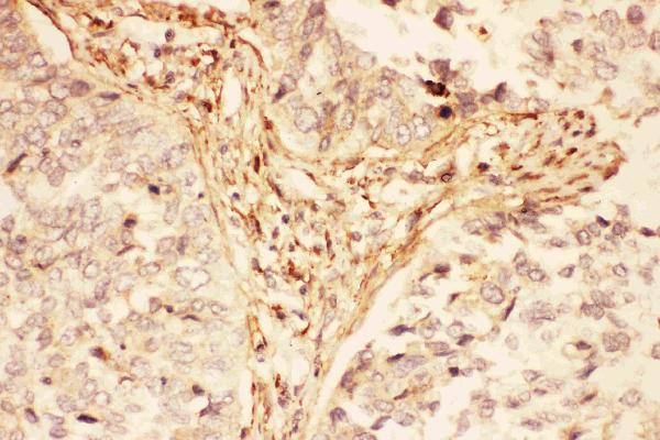
Figure 1. IHC analysis of LIF using anti-LIF antibody (PB9036).
LIF was detected in paraffin-embedded section of human lung cancer tissues. Heat mediated antigen retrieval was performed in citrate buffer (pH6, epitope retrieval solution) for 20 mins. The tissue section was blocked with 10% goat serum. The tissue section was then incubated with 1μg/ml rabbit anti-LIF Antibody (PB9036) overnight at 4°C. Biotinylated goat anti-rabbit IgG was used as secondary antibody and incubated for 30 minutes at 37°C. The tissue section was developed using Strepavidin-Biotin-Complex (SABC)(Catalog # SA1022) with DAB as the chromogen.

Click image to see more details
Figure 2. IHC analysis of LIF using anti-LIF antibody (PB9036).
LIF was detected in paraffin-embedded section of human placenta tissues. Heat mediated antigen retrieval was performed in citrate buffer (pH6, epitope retrieval solution) for 20 mins. The tissue section was blocked with 10% goat serum. The tissue section was then incubated with 1μg/ml rabbit anti-LIF Antibody (PB9036) overnight at 4°C. Biotinylated goat anti-rabbit IgG was used as secondary antibody and incubated for 30 minutes at 37°C. The tissue section was developed using Strepavidin-Biotin-Complex (SABC)(Catalog # SA1022) with DAB as the chromogen.

Click image to see more details
Figure 3. Western blot analysis of LIF using anti-LIF antibody (PB9036).
Electrophoresis was performed on a 5-20% SDS-PAGE gel at 70V (Stacking gel) / 90V (Resolving gel) for 2-3 hours. The sample well of each lane was loaded with 30 ug of sample under reducing conditions.
Lane 1: human Hela whole cell lysates,
Lane 2: human placenta tissue lysates,
Lane 3: human A431 whole cell lysates,
Lane 4: human A549 whole cell lysates.
After electrophoresis, proteins were transferred to a nitrocellulose membrane at 150 mA for 50-90 minutes. Blocked the membrane with 5% non-fat milk/TBS for 1.5 hour at RT. The membrane was incubated with rabbit anti-LIF antigen affinity purified polyclonal antibody (Catalog # PB9036) at 0.5 μg/mL overnight at 4°C, then washed with TBS-0.1%Tween 3 times with 5 minutes each and probed with a goat anti-rabbit IgG-HRP secondary antibody at a dilution of 1:5000 for 1.5 hour at RT. The signal is developed using an Enhanced Chemiluminescent detection (ECL) kit (Catalog # EK1002) with Tanon 5200 system. A specific band was detected for LIF at approximately 36 kDa. The expected band size for LIF is at 22 kDa.
Protein Target Info & Infographic
Gene/Protein Information For LIF (Source: Uniprot.org, NCBI)
Gene Name
LIF
Full Name
Leukemia inhibitory factor
Weight
22.008kDa
Superfamily
LIF/OSM family
Alternative Names
CDF; D Factor; DIA; differentiation inhibitory activity; differentiation stimulating factor; Differentiation-stimulating factor; Emfilermin; HILDA; HILDAcholinergic differentiation factor; leukemia inhibitory factor (cholinergic differentiation factor); leukemia inhibitory factor; LIF; Melanoma-derived LPL inhibitor; MLPLI LIF CDF, DIA, HILDA, MLPLI LIF interleukin 6 family cytokine leukemia inhibitory factor|D factor|cholinergic differentiation factor|differentiation inhibitory activity|differentiation-inducing factor|differentiation-stimulating factor|hepatocyte-stimulating factor III|human interleukin in DA cells|melanoma-derived LPL inhibitor
*If product is indicated to react with multiple species, protein info is based on the gene entry specified above in "Species".For more info on LIF, check out the LIF Infographic

We have 30,000+ of these available, one for each gene! Check them out.
In this infographic, you will see the following information for LIF: database IDs, superfamily, protein function, synonyms, molecular weight, chromosomal locations, tissues of expression, subcellular locations, post-translational modifications, and related diseases, research areas & pathways. If you want to see more information included, or would like to contribute to it and be acknowledged, please contact [email protected].
Specific Publications For Anti-LIF Antibody Picoband™ (PB9036)
Hello CJ!
PB9036 has been cited in 10 publications:
*The publications in this section are manually curated by our staff scientists. They may differ from Bioz's machine gathered results. Both are accurate. If you find a publication citing this product but is missing from this list, please let us know we will issue you a thank-you coupon.
LIF Upregulates Expression of NK-1R in NHBE Cells
Zhou L,Li C,Liu X,Zhang T.Effect of Irisin on LIF and integrin αvβ3 in rats of implantation failure.Reprod Biol Endocrinol.2021 Feb 3;19(1):18.doi:10.1186/s12958-021-00700-9.PMID:33536035;PMCID:PMC7856750.
Species: Rat
PB9036 usage in article: APP:IHC, SAMPLE:UTERINE TISSUE, DILUTION:NA
miR-182 aids in receptive endometrium development in dairy goats by down-regulating PTN expression
MicroRNA-223-3p suppresses leukemia inhibitory factor expression and pinopodes formation during embryo implantation in mice
Xu B, Sun X, Li L, Wu L, Zhang A, Feng Y. Fertil Steril. 2012 Aug;98(2):389-95. Doi: 10.1016/J.Fertnstert.2012.04.032. Epub 2012 Jun 13. Pinopodes, Leukemia Inhibitory Factor, Integrin-??3, And Mucin-1 Expression In The Peri-Implantation Endometri...
Pei K, Yu C, Shi X, Jia M. Contraception. 2010 Oct;82(4):379-84. Doi: 10.1016/J.Contraception.2010.04.009. Epub 2010 May 23. The Effects Of Mifepristone On The Expressions Of Osteopontin, Interleukin-6 And Leukemia Inhibitory Factor In The Villi O...
Li L, Xu Bf, Chen Qj, Sun Xx. Eur J Obstet Gynecol Reprod Biol. 2010 Aug;151(2):171-5. Doi: 10.1016/J.Ejogrb.2010.04.024. Epub 2010 Jun 9. Effects Of Hydrosalpinx On Pinopodes, Leukaemia Inhibitory Factor, Integrin Beta3 And Muc1 Expression In The...
Hu Cp, Feng Jt, Tang Yl, Zhu Jq, Lin Mj, Yu Me. Mediators Inflamm. 2006;2006(5):84829. Lif Upregulates Expression Of Nk-1R In Nhbe Cells.
Gui J, Xiong F, Yang W, Li J, Huang G. Am J Reprod Immunol. 2012 May;67(5):383-90. Doi: 10.1111/J.1600-0897.2011.01097.X. Epub 2012 Jan 9. Effects Of Acupuncture On Lif And Il-12 In Rats Of Implantation Failure.
Yu N, Yang J, Guo Y, Fang J, Yin T, Luo J, Li X, Li W, Zhao Q, Zou Y, Xu W. Am J Reprod Immunol. 2014 Jan;71(1):24-33. Doi: 10.1111/Aji.12150. Epub 2013 Aug 1. Intrauterine Administration Of Peripheral Blood Mononuclear Cells (Pbmcs) Improves Endo...
Recommended Resources
Here are featured tools and databases that you might find useful.
- Boster's Pathways Library
- Protein Databases
- Bioscience Research Protocol Resources
- Data Processing & Analysis Software
- Photo Editing Software
- Scientific Literature Resources
- Research Paper Management Tools
- Molecular Biology Software
- Primer Design Tools
- Bioinformatics Tools
- Phylogenetic Tree Analysis
Customer Reviews
Have you used Anti-LIF Antibody Picoband™?
Submit a review and receive an Amazon gift card.
- $30 for a review with an image
Be the first to review Anti-LIF Antibody Picoband™
*The first user to submit a review for a product is eligible for Boster's Innovating Scientists Reward, which gives product credits. This is in addition to the gift card reward.
Customer Q&As
Have a question?
Find answers in Q&As, reviews.
Can't find your answer?
Submit your question
5 Customer Q&As for Anti-LIF Antibody Picoband™
Question
We have tried in the past anti-LIF antibody for WB on left coronary artery in the past. I am using human, and We are going to use the antibody for IHC-P next. I would like examining left coronary artery as well as embryonic stem cell in our next experiment. Could give a recommendation on which antibody would work the best for IHC-P?
Verified Customer
Verified customer
Asked: 2020-04-10
Answer
I took a look at the website and datasheets of our anti-LIF antibody and I see that PB9036 has been validated on human in both WB and IHC-P. Thus PB9036 should work for your application. Our Boster satisfaction guarantee will cover this product for IHC-P in human even if the specific tissue type has not been validated. We do have a comprehensive range of products for IHC-P detection and you can check out our website bosterbio.com to find out more information about them.
Boster Scientific Support
Answered: 2020-04-10
Question
We are currently using anti-LIF antibody PB9036 for human tissue, and we are well pleased with the WB results. The species of reactivity given in the datasheet says human. Is it possible that the antibody can work on bovine tissues as well?
Verified Customer
Verified customer
Asked: 2020-02-12
Answer
The anti-LIF antibody (PB9036) has not been validated for cross reactivity specifically with bovine tissues, but there is a good chance of cross reactivity. We have an innovator award program that if you test this antibody and show it works in bovine you can get your next antibody for free. Please contact me if I can help you with anything.
Boster Scientific Support
Answered: 2020-02-12
Question
My question regards using your anti-LIF antibody for regulation of metanephric nephron tubule epithelial cell differentiation studies. Has this antibody been tested with western blotting on hela whole cell lysates? We would like to see some validation images before ordering.
Verified Customer
Verified customer
Asked: 2020-01-09
Answer
Thank you for your inquiry. This PB9036 anti-LIF antibody is tested on human placenta tissue, hela whole cell lysates, a549 whole cell lysates. It is guaranteed to work for IHC-P, WB in human. Our Boster guarantee will cover your intended experiment even if the sample type has not been be directly tested.
Boster Scientific Support
Answered: 2020-01-09
Question
We have seen staining in human colon. Are there any suggestions? Is anti-LIF antibody supposed to stain colon positively?
Verified Customer
Verified customer
Asked: 2019-08-21
Answer
Based on literature colon does express LIF. Based on Uniprot.org, LIF is expressed in left coronary artery, embryonic stem cell, colon, among other tissues. Regarding which tissues have LIF expression, here are a few articles citing expression in various tissues:
Colon, Pubmed ID: 15489334
Embryonic stem cell, Pubmed ID: 15146197
Boster Scientific Support
Answered: 2019-08-21
Question
My team were happy with the WB result of your anti-LIF antibody. However we have observed positive staining in colon secreted. using this antibody. Is that expected? Could you tell me where is LIF supposed to be expressed?
Verified Customer
Verified customer
Asked: 2019-08-09
Answer
According to literature, colon does express LIF. Generally LIF expresses in secreted. Regarding which tissues have LIF expression, here are a few articles citing expression in various tissues:
Colon, Pubmed ID: 15489334
Embryonic stem cell, Pubmed ID: 15146197
Boster Scientific Support
Answered: 2019-08-09
